Wednesday, January 30, 2019

Dental Implant Placement: An In-Depth Look at Precision Guided Surgery

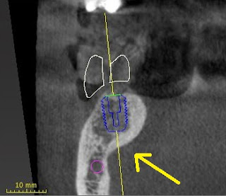
This latest blog will continue our discussion of using the latest CT digital imaging to plan and place dental implants.  Today's implant placement software allows us to not only see if an implant can be placed prior to surgery, but after placing a digital implant in the software, we can translate that information to your actual mouth and have the implant placed precisely where it was in the software. This removes almost all risk in implant placement error.  We also are excited to offer immediate implant placement following the same protocols.  Frequently patients come in on emergency with fractured or decayed teeth that cannot be restored and unfortunately have to be removed.  Immediate implants are dental implants placed the same day as taking the tooth out.  This dramatically cuts down on healing time and can decrease overall costs.  Prior to tooth removal, we can scan the mouth, digitally plan the case and have a custom guide fabricated for the day of surgery.  Statistically there is little difference in success rates of immediate vs. delayed implant placement as long as you follow strict protocols and proper surgical technique! Here is an image of the treatment planning software with an implant placed in a healed site:


As you can see, I even custom fabricated a digital tooth, so I can place the implant in the best possible position to support the future crown.  


This is what separates our surgical techniques from  other offices whom may place implants freehand with out any CT data or guide.  Our whole process and planning is based off the end result, a functional and aesthetic tooth.  

By using precision guided placement, a surgical guide stent is fabricated with a sleeve that only allows placement in the location that was predetermined in the implant planning software.  It is so accurate that the even the depth is predetermined and built into this surgical guide.


The implant placement is extremely predictable and rarely is there discomfort, especially if an implant is placed in a healed site. Here is an image of an implant placed using the above guide on the day of surgery with a custom healing cap:
 

After four to six months of healing, an impression is taken of the implant, and a custom abutment and crown are placed.  This new crown will function and feel like a natural tooth without the possibility of ever getting a cavity.
For more information, please visit our website: www.crowfamilydental.com

Thursday, July 30, 2015

CT Based Precision-Guided Dental Implant Placement

It is amazing how far technology has come in dentistry in a short period of time.  Today's blog is going to discuss placing implants using cutting edge Cone Beam Computed Tomography (also known as CBCT) imaging and guided implant placement computer software.   CT- based dental implant placement, I believe, will soon become the standard of care for implant surgery.  The benefits of having a CT done prior to implant placement are immense: 
  • We will know before surgery if you have enough bone to support an implant.  Traditional methods of determining bone quality with a standard x-ray only gives a two-dimensional image that shows only the height of bone. CBCT imaging gives a 3D view of the bone also allowing width and density measurements, to determine ultimate implant placement options. 
  • We can precisely locate vital structures such as nerves, adjacent teeth, bone defects, and bone topography.
  • We can use computers to precisely place the implant in not only the best location of bone quality, we can also place it in the best place for esthetics, and most importantly, function.  
  • By realizing the whole picture, seeing where the implant is going to go prior to surgery,  we have the ability to evaluate and discuss with our patients all risks, complications, and possible compromises that may be present in each individual case.  

Above is a screenshot of a treatment plan with multiple, fully adjustable windows giving every angle and view of the future implant site.  Each window can be opened and allows full rotation around the implant and surgical site.

Normal x-ray only showing height of bone
CT Image cross-section of same area of missing tooth
This is a classic example of why a CT image is so important!  There is no way to know looking at a normal radiograph that there is a major ledge and thinning of bone (indicated by the arrow).  This anatomical ledge greatly impacts placement and size/length of the dental implant.  You can imagine how valuable this information is for surgical procedures.  With improper technique and only basic x-rays, this case could end up with major problems if you thought there was plenty of height of bone for implant placement.  Our next blog will further discuss the surgical placement of implants using this newer technology.  For more information, please visit our website: www.crowfamilydental.com


Tuesday, December 23, 2014

Replacing Missing Teeth

When someone loses a tooth, it can lead to many problems.  Missing teeth can compromise aesthetics, speech, chewing, and increase the load on other teeth.
There used to be two options for replacement when someone would lose a tooth:

1) Fabricate a removable partial denture which can move and collect food underneath.


 2) Fabricate a fixed bridge which requires teeth on either side of the open space to be prepared for a connecting restoration with a fake tooth in the middle. Bridges are an excellent option for most cases but if one of the two teeth supporting the bridge goes bad, then the whole bridge could be at risk.


The newest option for replacing missing teeth is to surgically place a dental implant.


Actually, dental implants are becoming the standard of care for replacement of missing teeth when possible.  Implants are the most natural replacement of a missing tooth.  Benefits of implants compared to the other options include:  implants can't decay or get cavities, you can floss normally around an implant, there is no need for preparing the teeth around the open space.  The only requirement is sound bone and tissues to support the dental implant.  Implant placement has become very predictable and pain-free using the latest dental CT imaging and guided surgery.  Our future blog will discuss this newer procedure.  If you have any questions or would like more information, please visit us at www.crowfamilydental.com.

Thursday, June 5, 2014

The Greatest Game Changer in Dentistry: Cone Beam 3D CT Scan for Diagnosis and Treatment Planning

As most of you are aware, we have moved into a wonderful new facility located across the street from Walgreens in Park Ridge.  We invite you to take a tour of our new office via our website at crowfamilydental.com.  Unfortunately, our old Panorex x-ray machine was too old to make the move so we had to upgrade.  The piece of equipment we have in place now to replace our old machine is an Instrumentarium Orthopantomograph OP300. There are many exciting new features of this machine, a few of which we will be discussing in this blog.

Due to the latest digital technology, the OP300 significantly decreases the dosage and exposure to capture an image.  There is a significant reduction in dosage from our old machine compared to this current one. One full-mouth Panorex image on our new unit is equal in dosage to two normal digital x-rays.  The OP300 does an outstanding job capturing the whole mouth, sinuses, and temporomandibular joints in a computerized file format that allows excellent viewing and diagnosis.  There is a significant reduction in expense and waste by removing the need for film and processing chemicals.


I think the most exciting feature this new technology offers is that it captures cone beam 3D CT images.  By being able to rotate 360 degrees around a tooth, I have been able to see things going on that were never visible with standard x-rays. Here are some examples of some of the images we have taken so far.  On the left are traditional x-rays, and on the right are cross-sectional screen shots of the same teeth taken with this 3D CT technology.


By viewing only a traditional x-ray, we miss very valuable information which can affect the outcome of treatment.  The top left root canal-treated tooth could have been diagnosed for re-treatment based on this traditional x-ray.  The 3D CT image(on the upper right) saved a patient from a procedure that ultimately would have failed again due to a root defect.  The lower image shows a tooth that had a draining abscess and required root canal therapy.  The traditional x-ray on the lower left shows a single canal.  The 3D image on the right shows a canal/defect exiting the tooth at mid root and a very unique internal canal anatomy which would affect the outcome of treatment.  As you can imagine, this is a game changer for dentistry and our patients.  Our office is one of the first in Central Wisconsin to offer this low-dosage 3D cone beam CT technology.  One of these 3D scans is equivalent to a set of traditional Bitewing x-rays(4 films D-Speed).  This is also up to 50 times less dosage as compared to a medical CT of the head.  Our next blog will discuss using this technology for implant treatment planning and placement!  

Friday, September 27, 2013

Laser Periodontal Therapy

Crow Family Dental is proud to announce the certification of both of our dental hygienists.  In June, Terri and Janice completed course work in soft tissue management via the DEKA Laser, becoming some of the first hygienists in Wisconsin to earn this certificate!  Using a soft tissue laser for pocket decontamination and debridement offers many clinical benefits when added to conventional gum disease prevention and therapy.

Periodontal disease affects approximately 60% of adults and like heart disease and diabetes, without proper screening, you don't know there is a problem until it is too late.  Research has shown that gum disease is associated with other systemic health problems such as heart disease,  diabetes, and other chronic inflammatory conditions.  Bacteria are the enemy in our battle against periodontal disease.  We know that some of the bacteria do live in and around the surrounding tissues.  Therefore, cleaning only the surfaces of the teeth will leave behind the tissue-invasive bacteria which are free to produce and create new disease.  It is said that our bodies are not completely healthy until our gums are healthy.  With infected gums, your body's immune system has to work overtime; 24 hours a day.

The great thing about the laser is that it eliminates bacteria and their toxins that breakdown the bone that holds your teeth in place.  Laser therapy seals the blood vessels and nerve endings which eliminates the need for stitches and reduces swelling and inflammation.  The laser energy supports healing by stimulating your tissue with light energy to increase the oxygen supply and super charges the cells for faster healing.  The results of laser therapy have been unbelievable and patient satisfaction is outstanding.

Some of the benefits of laser therapy include:

  1. Efficiency & Precision
  2. Selective photo-thermal ablation
  3. Deeper penetration/aids in removing tarter
  4. Stimulates regeneration of tissue
  5. Neutralizes endotoxins/bactericidal
  6. Hemostasis/less sensitivity
  7. Quicker healing/minimally invasive for optimum patient comfort
  8. Reduction in bleeding gums, bacteria, pain and bad breath
  9. Teeth and smile are maintained
  10. No stitches are needed
  11. Minimal pain.
  12. Minimal downtime after treatment
    Minimal recession after treatment


If you have any questions about laser therapy, please visit us at www.crowfamilydental.com

Monday, March 4, 2013

Sleep Apnea and Snoring

Is snoring a problem in your home?  Are you tired of not getting a good night's sleep?

Snoring is an alarm that may be alerting you of a more serious problem known as Sleep Apnea.  Sleep apnea is a dangerous sleep disorder that occurs when a person's breathing is interrupted.  People with untreated sleep apnea stop breathing many times during their sleep, sometimes hundreds of times.  This means the brain and the rest of the body may not get enough oxygen therefore depriving your sleep and stressing your body.
There are two types of sleep apnea:
  • Obstructive Sleep Apnea:  This is the most common of the two forms of apnea, it is caused by a blockage of the airway, usually when the soft tissue in the back of the throat collapses during sleep.
  • Central Sleep Apnea: The airway is not blocked like in OSA, but the brain fails to signal the muscles to breathe due to instability in the respiratory control center.
Sleep apnea can affect anyone at any age, even children.  Some of the risk factors for sleep apnea include: 
  • being overwieght
  • being over age 40
  • having a larger neck size
  • having large tonsils
  • a large tongue
  • a small jaw bone
  • having a family history of sleep apnea
  • Gastroesophageal reflux
  • a nasal obstruction due to a deviated septum
  • allergies
  • sinus problems 
What are the effects of Sleep Apnea?  If Sleep Apnea is left untreated, it can result in a number of health problems such as:  High Blood Pressure, Stroke, Heart Failure, Irregular Heart Beats, and Heart Attacks, Diabetes, and Depression.  In addition, untreated sleep apnea may be responsible for poor performance in every day activities.  Problems with work or school, motor vehicle crashes, and academic underachievement in children and adolescents are just a few.
Common Sleep Apnea symptoms are waking up with a very sore throat, loud snoring, occasionally waking, sometimes waking with a choking or gasping sensation, sleepiness or lack of energy during the day, or sleepiness while driving.
What can be done if you suspect you are experiencing Sleep Apnea? Your dentist can help you!  Dr. Crow can screen you for sleep apnea with both a questionaire and home sleep testing monitor.  If you are at high risk, a sleep physician will further evaluate you for a final diagnosis and treatment.  Treatment could involve either a CPap Machine or an Oral Appliance fabricated by Dr. Crow.If you have any questions, please check out our website at www.crowfamilydental.com or give us a call!

Sleep Apnea Appliance


Thursday, January 17, 2013

Surprising Link of Sports and Energy Drinks to Tooth Decay

Did you know that sugar does not directly cause Cavities....It is actually acid that eats away your teeth that produces the cavities. The natural bacteria in your mouth and in plaque use sugar as food and the by product they produce is acid.  Unfortunately, a lot of foods and beverages we consume each day also bath our teeth in acid and we are all unaware of the potential damage we are doing!

Processed foods and beverages are littering the aisles of every grocery store, and more than likely every pantry across the country.  With this, the enamel of our nation’s youth is rapidly deteriorating.  Parents know they need to keep soda intake to a minimum in their children’s diets in order to avoid cavities and decay, but do they really know what kinds of choices are best for their children’s dental health?  What we don’t realize is that some of the most common beverages have such high levels of acid, resulting in a mouthful of cavities and irreversible damage to children’s teeth.

 Aside from these culprits, what other highly acidic beverages are bad for enamel?  We invite you to take a look at this infographic on pH levels of common beverages below.
 
 
Young adults consume these drinks with the assumption that they will improve their sports performance and energy levels.  They believe that these are better for them than "soda" and are shocked to find that these drinks are literally soaking their teeth in acid. 

Some research was done on the acidity levels of sports and energy drinks.  To test the effect of the acidity levels, the researchers immersed samples of human tooth enamel in each beverage for 15 minutes, followed by immersion in artificial saliva for two hours.  This cycle was repeated four times a day for five days, and the samples were stored in fresh artificial saliva at all other times.
This testing simulated the same exposure that a large proportion of American teens and young adults are subjecting their teeth to on a regular basis by drinking one of these beverages every few hours.
This research found that damage to enamel was evident after only five days of exposure to sports or energy drinks, although energy drinks showed a significantly greater potential to damage teeth than sports drinks.  In fact, energy drinks caused twice as much damage to teeth as sports drinks.  Damage caused to tooth enamel is irreversible.  Without the protection of enamel, teeth become overly sensitive,  more prone to cavities, and more likely to decay and wear.

It is recommended that  patients minimize their intake of sports and energy drinks.  It is also advised that they chew sugar-free gum or rinse their mouth with water following consumption of these drinks.  Both of these tactics increase saliva flow, which naturally helps to return the acidity levels in the mouth to normal.